Scalable volumetric imaging for ultrahigh-speed brain mapping at synaptic resolution
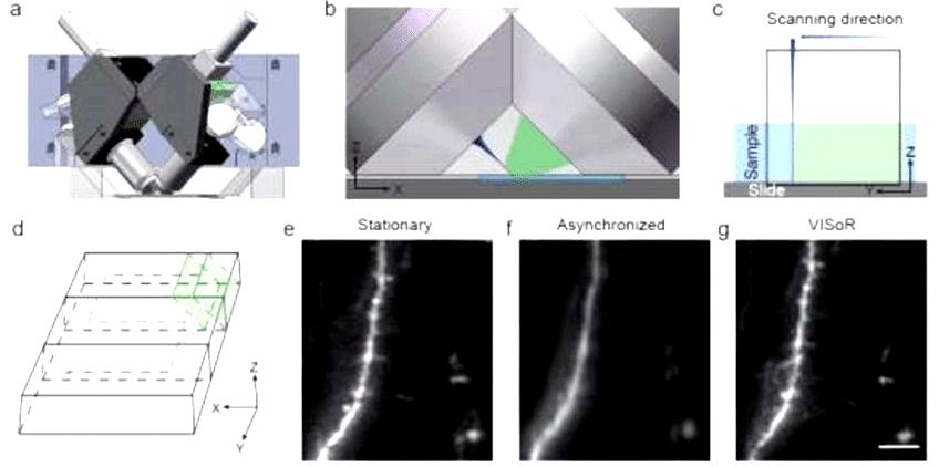
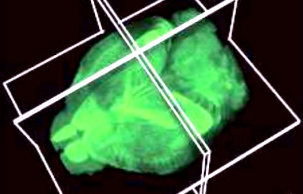
The rate of high definition optical imaging is a rate-restricting factor for meso-scale mapping of brain structures and functional circuits, that is of fundamental importance for neuroscience research. Here, we describe a brand new microscopy approach to Volumetric Imaging with Synchronized on-the-fly-scan and Readout (VISoR) for top throughput, top quality brain mapping. Mixing synchronized checking beam illumination and oblique imaging over removed tissue sections in smooth motion, the VISoR system effectively eliminates motion blur to acquire undistorted images. By continuously imaging moving samples without having to stop, the machine achieves high-speed 3D image purchase of a whole mouse brain within 1.5 hrs, in a resolution able to visualizing synaptic spines. A pipeline is produced for sample preparation, imaging, 3D image renovation and quantification. Our approach works with immunofluorescence methods, enabling flexible cell-type specific brain mapping, and it is readily scalable for big biological samples for example primate brains. By using this system, we examined behaviorally relevant whole-brain neuronal activation in 16 c-Fos-shEGFP rodents under resting or forced swimming conditions. Our results indicate the participation of multiple subcortical areas in stress response. Intriguingly, neuronal activation during these areas exhibits striking individual variability among different creatures, suggesting involve sufficient cohort size for such studies.

Resourse: https://academic.oup.com/nsr/advance-article-abstract/doi/10.1093/nsr/nwz053/


